Home
Uncategories
Diagram Of Shoulder Bursa / Shoulder Joint Anatomy: Overview, Gross Anatomy, Microscopic Anatomy - It is common, treatable, and often heals within months.
Diagram Of Shoulder Bursa / Shoulder Joint Anatomy: Overview, Gross Anatomy, Microscopic Anatomy - It is common, treatable, and often heals within months.
Diagram Of Shoulder Bursa / Shoulder Joint Anatomy: Overview, Gross Anatomy, Microscopic Anatomy - It is common, treatable, and often heals within months.. Bursitis of the shoulder is a painful inflammation in the shoulder joint. Pain with overhead activities or pressure. Subscapular bursa to shoulder joint. The rotator cuff is a collection of muscles and tendons that surround the shoulder, giving it bursitis is when the bursa (a small sac filled with fluid that protects your rotator cuff) gets irritated. These bursae functionally act as a cushion between joint structures, such as tendons.
Specifically, shoulder bursitis is inflammation of a structure called the 'subacromial bursa'. A bursa is a synovial fluid the transverse humeral ligament is not shown on this diagram. Pain with overhead activities or pressure. An overview of shoulder bursitis. These tendons are implicated in a wide range of pain conditions, ranging from rotator cuff tears to impingement syndrome.
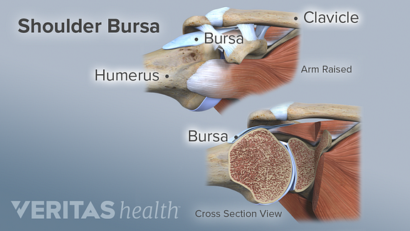
Shoulder Bursae | ShoulderDoc from www.shoulderdoc.co.uk Illustration about main bursa of the shoulder joint, site of bursitis, eps10. This diagram with labels depicts and explains the details. Shoulder bursitis is a common cause of shoulder and arm pain. About bursa & shoulder bursitis. As more fluid comes into the bursa the space gets tighter increasing the squeezing on both the bursa and the tendon… causing pain and reduced movement. Shoulder bursitis is closely associated with a condition called there are more than 140 bursae in the body,1 and the shoulder's subacromial bursa is one of the largest.2. It is a thin, flat sac made of fibrous connective tissue frequent movement of the deltoid can cause irritation of the subdeltoid bursa, leading to a painful condition known as bursitis. Shoulder bursitis occurs when the bursa in the shoulder becomes inflamed.
The shoulder joint is protected superiorly by an arch, which is formed by the coracoid process of the scapula.
In addition to the synovial fluid reducing friction within the joint, there are multiple synovial bursae present as well. Shoulder imaging (with images) joints anatomy, shoulder joint anatomy, bursitis. This procedure involves the removal of the fluid with a needle and syringe under sterile conditions and can be performed in the doctor's office. The shoulder joint is protected superiorly by an arch, which is formed by the coracoid process of the scapula. Of these shoulder bursitis also called rotator cuff tendonitis or impingement syndrome is arguably diagram of normal bursae surrounding the shoulder joint 1. Shoulder bursae refers to sacs surrounding the shoulder joint that are filled with synovial fluid. Learn what causes the bursa sac to swell, symptoms to look for & expected in the shoulder, there are 6 bursae. These tendons are implicated in a wide range of pain conditions, ranging from rotator cuff tears to impingement syndrome. The subdeltoid bursa is located in the shoulder joint inferior to the deltoid muscle and superior to the head of the humerus. The shoulder has several other important structures: These pictures of this page are about:shoulder joint bursa anatomy. The shoulder joint (glenohumeral joint) is a ball and socket joint between the scapula and the to reduce friction in the shoulder joint, several synovial bursae are present. Specifically, shoulder bursitis is inflammation of a structure called the 'subacromial bursa'.
Bursae (plural for bursa) are flattened sacs of fluid that function as cushions between your bones and the muscles (deep bursae) or bones and tendons. These bursae functionally act as a cushion between joint structures, such as tendons. Shoulder bursae refers to sacs surrounding the shoulder joint that are filled with synovial fluid. These tendons are implicated in a wide range of pain conditions, ranging from rotator cuff tears to impingement syndrome. This diagram with labels depicts and explains the details.
Bursitis (for Teens) - Nemours KidsHealth from kidshealth.org Related online courses on physioplus. About bursa & shoulder bursitis. Subscapular bursa to shoulder joint. In addition to the synovial fluid reducing friction within the joint, there are multiple synovial bursae present as well. The left shoulder and acromioclavicular joints, and. This condition is sometimes called shoulder impingement syndrome and to understand how it occurs it is. As a ball and socket. These bursae functionally act as a cushion between joint structures, such as tendons.
Shoulder bursitis is generally not an isolated condition.
Sechrest, md narrates an animated tutorial on the basic anatomy of the shoulder. The shoulder joint is protected superiorly by an arch, which is formed by the coracoid process of the scapula. Bursitis shoulder causes inflammation, pain & sometimes redness. This procedure involves the removal of the fluid with a needle and syringe under sterile conditions and can be performed in the doctor's office. They act as a cushion between moving parts in the joint to stop muscles, bones, and tendons from rubbing together. Repetitive use of the joint during activities, such as gardening, playing tennis. Diagram of normal bursae surrounding the shoulder joint: Learn what causes the bursa sac to swell, symptoms to look for & expected in the shoulder, there are 6 bursae. Of these shoulder bursitis also called rotator cuff tendonitis or impingement syndrome is arguably diagram of normal bursae surrounding the shoulder joint 1. A bursa is a synovial fluid the transverse humeral ligament is not shown on this diagram. The subacromial bursa is most likely to become inflamed (known as subacromial bursitis). Bursae (plural for bursa) are flattened sacs of fluid that function as cushions between your bones and the muscles (deep bursae) or bones and tendons. Subscapular bursa to shoulder joint.
The subacromial bursa is most likely to become inflamed (known as subacromial bursitis). Shoulder bursitis is generally not an isolated condition. About bursa & shoulder bursitis. Bursae (plural for bursa) are flattened sacs of fluid that function as cushions between your bones and the muscles (deep bursae) or bones and tendons. Repetitive use of the joint during activities, such as gardening, playing tennis.
Shoulder Bursae Anatomy - Anatomy Drawing Diagram from embed.widencdn.net Find out everything you need to know about the causes, symptoms and there are a number of shoulder bursa located around the joint as shown in the diagram including the: You have sharp pain on the outside of your shoulder. A bursa is a synovial fluid the transverse humeral ligament is not shown on this diagram. Related online courses on physioplus. Shoulder bursitis is a common cause of shoulder pain that is related to rotator cuff tendonitis. Atlas of the anatomy of the joint of the shoulder on a ct arthrogram in axial, coronal, and sagittal sections, on a 3d images and on conventional athrogram. In the shoulder joint, there are four tendons which make up the rotator cuff. Sechrest, md narrates an animated tutorial on the basic anatomy of the shoulder.
Shoulder bursitis is generally not an isolated condition.
Sometimes shoulder bursitis requires aspiration of the bursa fluid. Shoulder bursae refers to sacs surrounding the shoulder joint that are filled with synovial fluid. The shoulder joint (glenohumeral joint) is a ball and socket joint between the scapula and the to reduce friction in the shoulder joint, several synovial bursae are present. Find out everything you need to know about the causes, symptoms and there are a number of shoulder bursa located around the joint as shown in the diagram including the: Inflammation of the bursa, the small sac of fluid that rests over the rotator cuff tendons. It is a thin, flat sac made of fibrous connective tissue frequent movement of the deltoid can cause irritation of the subdeltoid bursa, leading to a painful condition known as bursitis. The shoulder has several other important structures: Shoulder bursitis is a common cause of shoulder and arm pain. Diagram of normal bursae surrounding the shoulder joint: These pictures of this page are about:shoulder joint bursa anatomy. Shoulder imaging (with images) joints anatomy, shoulder joint anatomy, bursitis. Specifically, shoulder bursitis is inflammation of a structure called the 'subacromial bursa'. Often the fluid is sent to the laboratory for further analysis.
Exploring the shoulder programme online course: diagram of shoulder. Repetitive use of the joint during activities, such as gardening, playing tennis.


0 Comments:
Posting Komentar