Home
Uncategories
Diagram Of Hip.and Back.muscles / Muscles Move And Support The Spine - Piriformis, external and internal obturators and the superior and inferior gemelli.
Diagram Of Hip.and Back.muscles / Muscles Move And Support The Spine - Piriformis, external and internal obturators and the superior and inferior gemelli.
Diagram Of Hip.and Back.muscles / Muscles Move And Support The Spine - Piriformis, external and internal obturators and the superior and inferior gemelli.. The hip joint is made up of two. The muscles of the back are a group of strong, paired muscles that lie on the posterior aspect of the trunk they provide movements of the spine, stability to the trunk, as well as the coordination between the movements of the limbs and the back muscles are divided into two large groups: The hip abductors consist of the: These muscles can be grouped based upon their location and function. To learn more about the anatomy of the spine, watch this video.
The pubis, ischium, and ilium together constitute the pelvis while the thigh bone is the femur. The muscles of the lower back help stabilize, rotate, flex, and extend the spinal column, which is a bony tower of 24 vertebrae that gives the body structure and houses the spinal cord. To learn more about the anatomy of the spine, watch this video. The psoas muscle is a deep muscle that connects your spine to your leg.in fact, it's the only muscle that does so. This video also provides you with a.
Muscle Strains It Band Groin Hip Flexor Sports Medicine from sportsmedicine.mayoclinic.org The muscles of the lower back help stabilize, rotate, flex, and extend the spinal column, which is a bony tower of 24 vertebrae that gives the body structure and houses the spinal cord. Nerves in your lower back. The iliacus originates on the iliac fossa of the ilium. The hip abductors consist of the: The bones of the hip include the femur, the ilium, the ischium, and the pubis. Three types of back muscles that help the spine function are extensors, flexors and obliques. The muscles of the abdomen, lower back, and pelvis are separated from those of the chest by the muscular wall of the diaphragm, the critical breathing muscle. The four groups are the anterior group, the posterior group, adductor group, and finally the abductor group.
The iliacus originates on the iliac fossa of the ilium.
The hip itself is a ball and socket joint, much like the shoulder.the structures necessary to create this joint are the socket, the joint capsule, muscle, ligaments, and the neck. Muscle anatomy drawing 12 photos of the muscle anatomy drawing anatomy muscle sketches, arm muscle anatomy drawing, back muscle anatomy drawing, human muscle anatomy drawing, muscle anatomy drawing, human muscles, anatomy muscle sketches, arm muscle anatomy drawing, back muscle anatomy drawing, human muscle. The trapezius is a broad, flat and triangular muscle. Nerves carry signals from the brain to the muscles to move the hip and carry signals from the muscles back to the brain about pain, pressure and temperature. See back muscles and low back pain. The sciatic nerve is the most commonly recognized nerve in the hip and thigh. Most of the time, back muscle pain is diagnosed then treated with little more than a prescription of rest, painkillers and muscle relaxants. The bones of the hip include the femur, the ilium, the ischium, and the pubis. The muscles of the back are a group of strong, paired muscles that lie on the posterior aspect of the trunk they provide movements of the spine, stability to the trunk, as well as the coordination between the movements of the limbs and the back muscles are divided into two large groups: The muscles of the abdomen, lower back, and pelvis are separated from those of the chest by the muscular wall of the diaphragm, the critical breathing muscle. The four groups are the anterior group, the posterior group, adductor group, and finally the abductor group. Related posts of lower back muscles diagram muscle anatomy of the thigh. Three types of back muscles that help the spine function are extensors, flexors and obliques.
Muscles located at the side of the hip, which include the gluteus medius, piriformis, and hip external rotator muscles contribute greatly to the well being of your lower back, as well as your posture.when these muscles get tight, as they often do, you may find that along with hip pain, your lower back hurts—but you can't figure out why. Lower back muscle diagram anatomy The pelvic floor muscles provide foundational support for the intestines and bladder. The extensor muscles are attached to back of the spine and enable standing and lifting objects. Most of the time, back muscle pain is diagnosed then treated with little more than a prescription of rest, painkillers and muscle relaxants.
Crossfit Hip Musculature Part 2 Posterior Muscles from www.crossfit.com Muscle anatomy drawing 12 photos of the muscle anatomy drawing anatomy muscle sketches, arm muscle anatomy drawing, back muscle anatomy drawing, human muscle anatomy drawing, muscle anatomy drawing, human muscles, anatomy muscle sketches, arm muscle anatomy drawing, back muscle anatomy drawing, human muscle. The bones together make up the hip. The fibres attach to the clavicle, acromion and the scapula spine. These muscles can be grouped based upon their location and function. The muscles on each side form a trapezoid shape. Muscles located at the side of the hip, which include the gluteus medius, piriformis, and hip external rotator muscles contribute greatly to the well being of your lower back, as well as your posture.when these muscles get tight, as they often do, you may find that along with hip pain, your lower back hurts—but you can't figure out why. The muscles of the abdomen, lower back, and pelvis are separated from those of the chest by the muscular wall of the diaphragm, the critical breathing muscle. The pubis, ischium, and ilium together constitute the pelvis while the thigh bone is the femur.
Muscles located at the side of the hip, which include the gluteus medius, piriformis, and hip external rotator muscles contribute greatly to the well being of your lower back, as well as your posture.when these muscles get tight, as they often do, you may find that along with hip pain, your lower back hurts—but you can't figure out why.
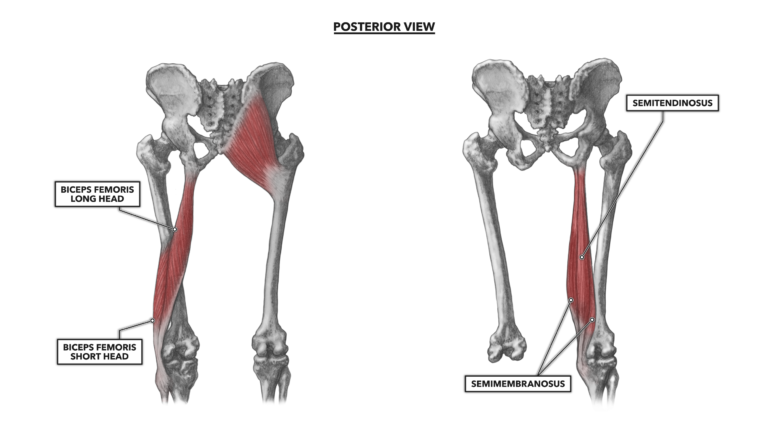
The muscles of the back are a group of strong, paired muscles that lie on the posterior aspect of the trunk they provide movements of the spine, stability to the trunk, as well as the coordination between the movements of the limbs and the back muscles are divided into two large groups: It runs from your lower back through your pelvis, passing to the front of your hip where it attaches to the top of your femur, which is your thigh bone. The iliacus originates on the iliac fossa of the ilium. The muscles on each side form a trapezoid shape. The bones of the hip include the femur, the ilium, the ischium, and the pubis. Muscle anatomy of the thigh 12 photos of the muscle anatomy of the thigh anatomy of the anterior thigh muscles, muscle anatomy of upper thigh, muscle anatomy thigh mri, muscles of the leg grey's anatomy, muscles of the thigh ct anatomy, human muscles, anatomy of the anterior thigh muscles, muscle anatomy of upper. Muscle anatomy drawing 12 photos of the muscle anatomy drawing anatomy muscle sketches, arm muscle anatomy drawing, back muscle anatomy drawing, human muscle anatomy drawing, muscle anatomy drawing, human muscles, anatomy muscle sketches, arm muscle anatomy drawing, back muscle anatomy drawing, human muscle. The psoas muscle is a deep muscle that connects your spine to your leg.in fact, it's the only muscle that does so. The fibres attach to the clavicle, acromion and the scapula spine. Gluteus maximus trigger point pain is felt toward the back of the hip and thigh near the hip joint, the base of the spine, and in the upper buttock going down alongside and into the gluteal fold. Piriformis, external and internal obturators and the superior and inferior gemelli. The four groups are the anterior group, the posterior group, adductor group, and finally the abductor group. Sprains and strains are a common cause of pain around the back and hips.
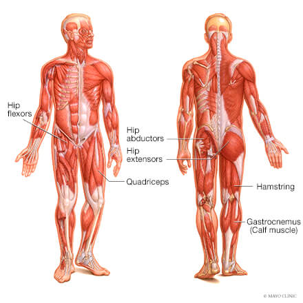
Like the forearm, the upper leg, or thigh, has a dense arrangement of many muscles. To learn more about the anatomy of the spine, watch this video. If you are starting to feel hip pain or stiffness, you'll want to know more about the bones and muscles that make up the hip's anatomy. Nerves in your lower back. The psoas major is a large muscle that runs from the bodies and disc of the l1 to l5 vertebrae, joins with the iliacus via its tendon, and connects to the lesser trochanter of the femur.
What Is Piriformis Syndrome Causes Symptoms And Treatment from stretchcoach.com In physical therapy, a therapist will determine if you need to stretch the lower back muscles and other muscles such as the piriformis or hamstrings. It is the most superficial of all the back muscles. Related posts of muscles of the lower back and hip diagram muscle anatomy drawing. Nerves carry signals from the brain to the muscles to move the hip and carry signals from the muscles back to the brain about pain, pressure and temperature. On the anterior side, the most prominent of the muscles are the sartorius muscle and the four muscles that make up quadriceps muscle group (the quads.) Lower back muscle diagram anatomy The piriformis is the horizontal muscle in the center of the picture running over the top of the sciatic nerve. This is a diagram of the larger and more surface muscles of the low back.
The sciatic nerve is the most commonly recognized nerve in the hip and thigh.
The iliacus and psoas major comprise the iliopsoas group. The hip abductors consist of the: As you can see from the diagram to the right, there are many muscles and tendons that make up the hip and buttocks region. The pain will occasionally descend into the upper thigh. Three types of back muscles that help the spine function are extensors, flexors and obliques. Nerves in your lower back. The bones of the hip include the femur, the ilium, the ischium, and the pubis. Related posts of lower back muscles diagram muscle anatomy of the thigh. It runs from your lower back through your pelvis, passing to the front of your hip where it attaches to the top of your femur, which is your thigh bone. Like the forearm, the upper leg, or thigh, has a dense arrangement of many muscles. The sciatic nerve is the most commonly recognized nerve in the hip and thigh. The pelvic floor muscles provide foundational support for the intestines and bladder. A sprain is a torn or overstretched ligament, while a strain is a torn or overstretched tendon or muscle.

0 Comments:
Posting Komentar